


7月4日下午,中盛溯源生物科技有限公司组织了关于“公司风险管理之保密管理”的培训会,由我公司法律顾问周志芳为大家讲解保密工作的重要意义。公司全体员工悉数与会。
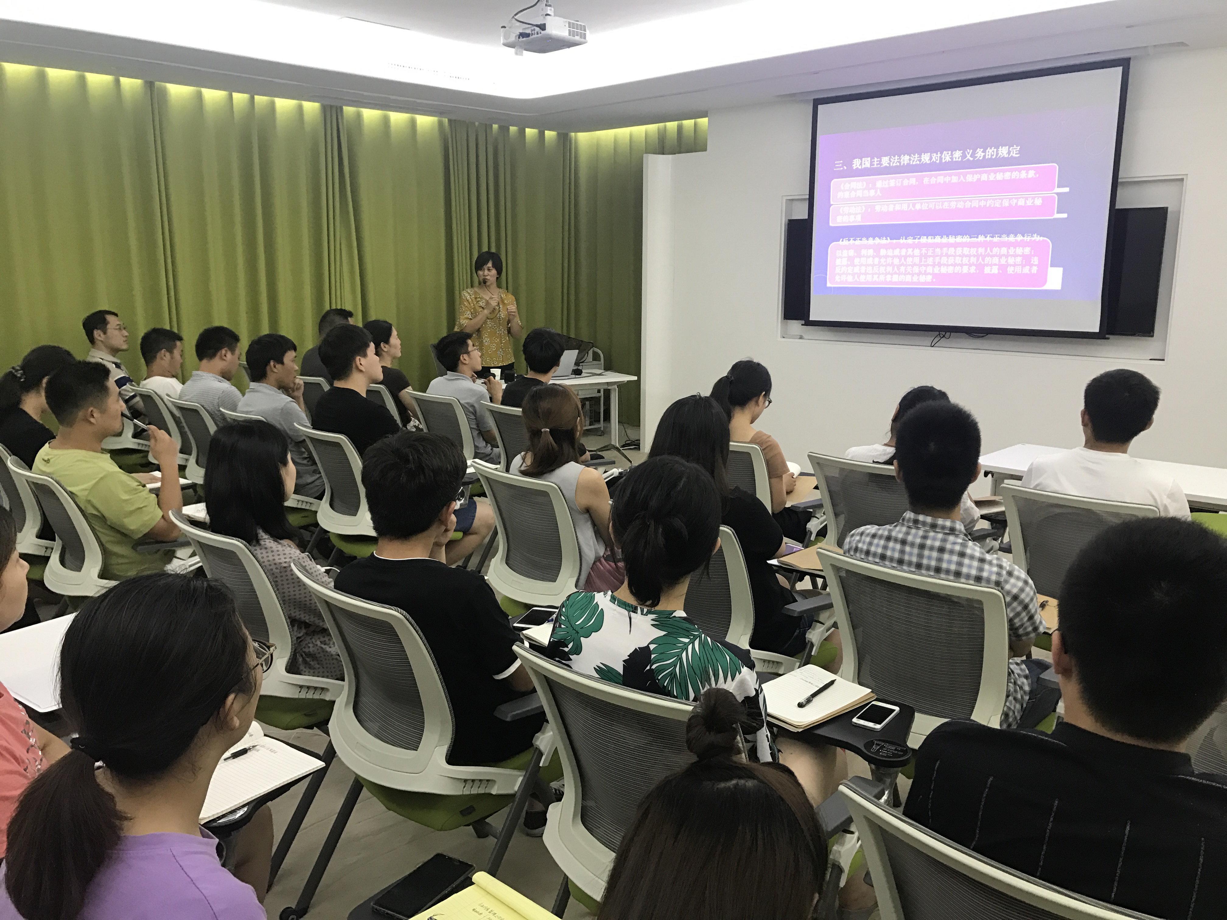
培训会上,周律师深刻分析了新形势下保密工作面临的复杂形势和严峻挑战,并强调保密工作无小事,泄密不仅影响个人前途,甚至给公司造成无法挽回的损失。期间,周律师特别着重讲解了信息安全的实际风险,并举例相关事实案件,加以深刻印象。
培训会图文并茂、案例详实,既有专业性的讲授,又有操作性的指导,紧扣保密工作主题,使参训人员了解了日常工作中有意无意地泄密行为带来的危害,加深了对保密工作相关的法律条规理解,提高了保密意识,增强了公司风险管控能力。

 Back
Back